Fetal morphology

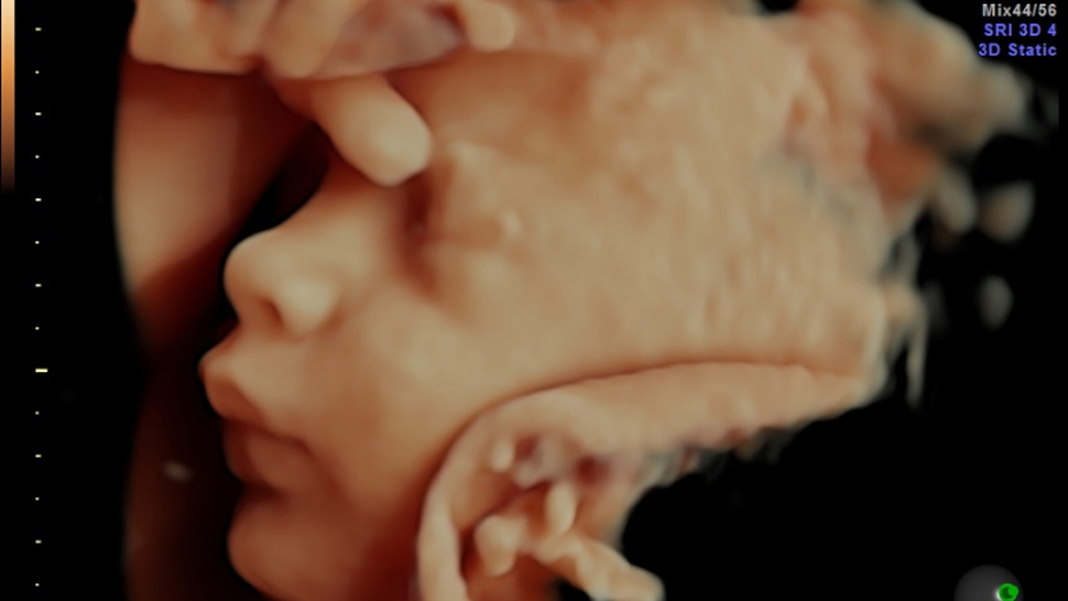
Fetal morphology is a highly specialized analysis performed with the help of an ultrasound. The goal of the procedure is to assess the anatomical development of the fetus throughout the different stages of pregnancy, namely the first, second and third trimesters.
The method is not dangerous for either the mother, nor the baby, and it can be part of any routine ultrasound examination. Our obstetrician-gynaecologists are certified to carry out fetal morphology as a diagnostic method, as it is an essential part of the proper follow-up of a pregnancy.
The main goal of the examination is the early diagnosis of moderate and sever anatomical discrepancies, incompatible with life and social adaptation. Hence, Saint Lazar Maternity Hospital is equipped with ultrasound apparatuses of the highest quality, which allows an extremely detailed examination of the fetus at every point of development.
It is of extreme importance, that fetal morphology is performed by highly qualified specialists, especially when it comes to prenatal diagnosis. During each stage of pregnancy, the examination is performed at a different scale. We recommend at least two follow-up examinations with a fetal morphology scan are performed during pregnancy.
Examination of the morphology of the fetus is performed during three main periods of its’ growth, during each trimester, via ultrasound examination:
First trimester (FM performed between the 11th and 13th gestational weeks):
During the first trimester and when there is normal growth of the fetus, we can visualize the development of the limbs, major blood vessels, the stomach, the urinary bladder, and the heart.
Early fetal morphology examinations can show various genetic abnormalities and can direct us towards laboratory test for chromosomal anomalies. The clearly depicted nasal bone and the thickness of the nuchal translucency allow the specialist to confirm or deny doubts for Down syndrome.
Second trimester (FM performed between the 18th and 23rd gestational weeks):
During the second fetal morphology scan the fetus begins to move, to hear and to open his/her eyes.
What do we see during the second fetal scan?
- A well-formed face
- Well-developing brain and bones of the skull
- Development of the thoracic cage
- Development of heart
- If any defects of the abdominal wall and urinary tract have appeared
- Development of the genitalia and blood vessels
Third trimester (FM performed between the 32nd and 34th gestational weeks):
During the third fetal morphology scan, we can obtain information about the general organ systems of the baby:
- Status of the central nervous system
- Cardiac status
- Well-developing gastrointestinal tract
- Well-developing urinary tract
- Well-developing limbs
Do not hesitate to book your fetal morphology scan with our certified specialists, namely Dr. Valentin Nikolov, Dr. Tiyana Nikolova and Dr. Veselina Slavkova!

